Upper Thigh Anatomy - How Do The Anatomy Of Knee And Lower Leg Affect Movement : Abductors are located on the upper portion of the outside of your thighs and hips, anchoring above on the pelvis, and below at various points on your outside thigh.
Upper Thigh Anatomy - How Do The Anatomy Of Knee And Lower Leg Affect Movement : Abductors are located on the upper portion of the outside of your thighs and hips, anchoring above on the pelvis, and below at various points on your outside thigh.. The head of the femur joins the pelvis (acetabulum) to form the hip joint. Upper thigh muscle anatomy : Legs give us the freedom to run, walk, jump, climb, and negotiate the world around us. Spicermanyt at checkout for 40% off this tutorial! 5.0 based on 12 ratings, 7 reviews.
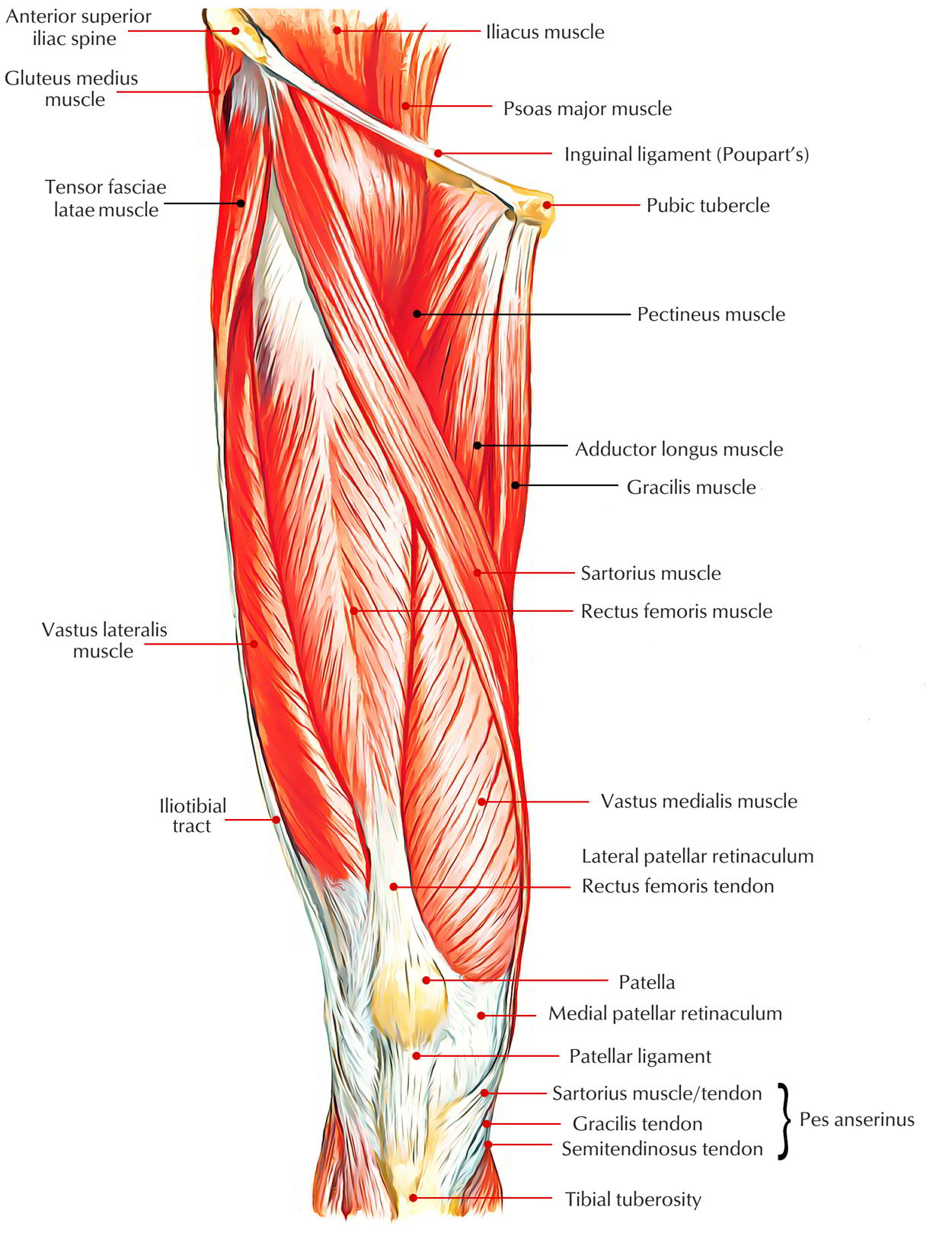
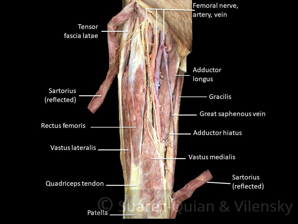
It contains many muscles and nerves but only has one bone, the femur, which is the longest and strongest bone in. Meanwhile, the vastus lateralis is on the side of the thigh, while the vastus intermedius is hidden below the rectus femoris(5). Like the adductors, the abductors are also responsible for stabilizing your knees during athletic and everyday movement. The iliacus muscle continues down through the pelvis and attaches to the small piece of bone (lesser trochanter) that is attached to your femur (upper thigh bone). This mri brain cross sectional anatomy tool is absolutely free to use.
Easy Notes On Muscles Of Anterior Compartment Of The Thigh Earth S Lab from www.earthslab.com 5.0 based on 12 ratings, 7 reviews. The thigh bears much of the load of the body's weight when a person is upright. The upper part of the thigh bone consists of the femoral head, femoral neck, and greater and lesser trochanters. The four muscles all extend the lower leg. The hamstring portion of the adductor magnus has a similar action to these muscles, but is located in the medial thigh. The thigh muscles don't just move your legs. This mri brain cross sectional anatomy tool is absolutely free to use. Rectus femoris muscle, one of the quadriceps muscles on the front of your thigh.
The vastus lateralis is a muscle located on the lateral, or outside, part of your thigh.
Check spelling or type a new query. The head of the femur joins the pelvis (acetabulum) to form the hip joint. They have a lot to do with how your hips move. The thigh muscles don't just move your legs. The upper thigh muscle pain or we can say in other words that is the discomfort in your upper thigh, this pain is very popular. On the anterior side, the most prominent of the muscles are the sartorius muscle and the four muscles that make up quadriceps muscle group (the quads.) The posterior upper leg muscles provide your knees with mobility (extension, flexion and rotation) and strength. The rectus femoris is located in the center of the thigh, while the vastus medialis is in the middle of the said body part. Read on for more information on causes and treatment options. Next to the femoral neck, there are two protrusions known as greater and lesser trochanters which serve as sites of muscle attachment. A deep, shooting pain in the upper leg can also be caused by deep vein thrombosis, spinal stenosis, or a thigh bone infection. Rectus femoris muscle, one of the quadriceps muscles on the front of your thigh. This webpage presents the anatomical structures found on thigh mri.
The iliacus muscle continues down through the pelvis and attaches to the small piece of bone (lesser trochanter) that is attached to your femur (upper thigh bone). Upper thigh muscle anatomy : It contains many muscles and nerves but only has one bone, the femur, which is the longest and strongest bone in. On the anterior side, the most prominent of the muscles are the sartorius muscle and the four muscles that make up quadriceps muscle group (the quads.) Take the upper extremity anatomy quiz and learn more about the bones, joints, muscles and vessels of the upper extremity!
Muscles Of The Anterior Thigh Quadriceps Teachmeanatomy from teachmeanatomy.info The upper thigh muscle pain or we can say in other words that is the discomfort in your upper thigh, this pain is very popular. The vastus lateralis is a muscle located on the lateral, or outside, part of your thigh. The vastus laterails works with the other quad muscles to help extend your knee joint. Upper thigh muscle pain is a very hard pain we can feel and didn't know how to treat it. This mri brain cross sectional anatomy tool is absolutely free to use. It contains many muscles and nerves but only has one bone, the femur, which is the longest and strongest bone in. Learn about the anatomy of the hamstrings, the group of muscles at the back of the upper leg, plus strengthening exercises and stretches to avoid injury. Most people can get pain in this area.
It's the area that runs from the hip to the knee in each leg.
Anterior muscles extend your legs and flex your thighs. Abductors are located on the upper portion of the outside of your thighs and hips, anchoring above on the pelvis, and below at various points on your outside thigh. Upper thigh muscle pain is a very hard pain we can feel and didn't know how to treat it. Maybe you would like to learn more about one of these?. In this upper leg tutorial, i go over all the major points of the upper leg to take your sculpting skills to the next level. Next to the femoral neck, there are two protrusions known as greater and lesser trochanters which serve as sites of muscle attachment. It also is active in maintaining thigh and kneecap position. Medial muscles adduct and rotate your thigh, and posterior flex your leg and extend your thigh. Human muscle anatomy 12 photos of the human muscle anatomy human anatomy muscle questions, human anatomy muscles clay learning system, human muscle anatomy head, human muscle anatomy leg, human muscle anatomy worksheet, human muscles, human anatomy muscle questions, human anatomy muscles clay learning system, human muscle. One further muscle of the anterior knee is the small articularis genus muscle, it is occasionally is blended with vastus intermedius. Knee assessment and hip mechanics online course: On the anterior side, the most prominent of the muscles are the sartorius muscle and the four muscles that make up quadriceps muscle group (the quads.) Most people can get pain in this area.
Upper thigh anatomy (page 1). This mri brain cross sectional anatomy tool is absolutely free to use. Abductors are located on the upper portion of the outside of your thighs and hips, anchoring above on the pelvis, and below at various points on your outside thigh. Upper inner thigh anatomy : The vastus laterails works with the other quad muscles to help extend your knee joint.
Sartorius Muscle Wikipedia from upload.wikimedia.org The head of the femur joins the pelvis (acetabulum) to form the hip joint. They have a lot to do with how your hips move. Spicermanyt at checkout for 40% off this tutorial! Muscles of the leg and foot classic human anatomy in motion the artist s guide to the dynamics of figure drawing : The rectus femoris is located in the center of the thigh, while the vastus medialis is in the middle of the said body part. Upper inner thigh anatomy : On the other hand, this pain is common. Severe leg pain located around the thigh can be caused by trauma from a femoral break or muscle strain.
Read on for more information on causes and treatment options.
Human muscle anatomy 12 photos of the human muscle anatomy human anatomy muscle questions, human anatomy muscles clay learning system, human muscle anatomy head, human muscle anatomy leg, human muscle anatomy worksheet, human muscles, human anatomy muscle questions, human anatomy muscles clay learning system, human muscle. The muscles in the upper leg power many of our movements. On the anterior side, the most prominent of the muscles are the sartorius muscle and the four muscles that make up quadriceps muscle group (the quads.) By spicer mcleroy in tutorials. Spicermanyt at checkout for 40% off this tutorial! On the other hand, this pain is common. Anterior muscles extend your legs and flex your thighs. Next to the femoral neck, there are two protrusions known as greater and lesser trochanters which serve as sites of muscle attachment. The upper part of the thigh bone consists of the femoral head, femoral neck, and greater and lesser trochanters. The hamstring portion of the adductor magnus has a similar action to these muscles, but is located in the medial thigh. Rectus femoris muscle, one of the quadriceps muscles on the front of your thigh. Muscles play an important role in the. The adductor brevis, adductor longus and adductor magnus make up the the starting position is lying on the right side where the upper body is supported by the right arm.


0 Komentar